Tampilkan postingan dengan label BAKTERIOLOGI 3. Tampilkan semua postingan

Rabu, 12 September 2018

STERILISASI


Sterilisasi dalam mikrobiologi adalah suatu proses untuk mematikan semua organisme yang terdapat pada atau di dalam suatu benda. Ketika untuk pertama kalinya melakukan pemindahan biakan bakteri secara aseptik, sesungguhnya hal itu telah menggunakan salah satu cara sterilisasi, yaitu pembakaran. Namun, kebanyakan peralatan dan media yang umum dipakai di dalam pekerjaan mikrobiologi akan menjadi rusak bila dibakar. Ada tiga cara utama yang umum dipakai dalam sterilisasi yaitu penggunaan panas, bahan kimia, dan penyaringan atau filtrasi 
Sterilisasi yang baik dapat mencegah tumbuhnya mikroba lain yang tidak diharapkan dalam bahan yang telah disterilisasi. Teknik sterilisasi yang digunakan berbeda antara satu dengan lainnya, tergantung dari jenis material yang digunakan. Alat-alat yang digunakan dalam praktikum mikrobiologi juga harus dalam keadaan steril atau bebas dari kuman serta bakteri, virus dan jamur. Dan untuk mensterilkannya diperlukan pula pengetahuan tentang cara-cara dan teknik sterilisasi. Hal ini dilakukan karena alat- alat yang digunakan pada laboratorium mikrobiologi memiliki teknik sterilisasi yang berbeda 
Steril merupakan syarat mutlak keberhasilan kerja dalam laboratorium mikrobiologi. Dalam melakukan sterilisasi, diperlukan teknik-teknik agar sterilisasi dapat dilakukan secara sempurna, dalam arti tidak ada mikroorganisme lain yang  mengkontaminasi media. Ada beberapa teknik sterilisasi, yaitu dengan cara fisikdengan panas, mekanik dengan filtrasi dan kimia dengan senyawa-senyawa kimia. Pembersihan benda-benda atau permukaan tubuh akan mengurangi jumlah mikroba sehingga memperkecil kemungkinan terjadinya infeksi. Misalnya cuci tangan dengan sabun dan bilas dengan air mengalir sebelum mengoperasikan 
Pada pengerjaan mikrobiologi, diperlukan suatu kondisi yang benar-benar aseptik dimana alat penunjang serta nutrient dan substrat harus benar-benar steril. Hal ini berarti mikroba kontaminan harus dimatikan. Sterilisasi dilakukan pada suhu 121oC selama 30 menit, yaitu agar spora atau mikroba dapat dimatikan. Spora adalah sel istirahat yang resisitan terhadap panas dan lingkungan yang berfungsi sebagai tunas untuk berkembang biak selanjutnya. Udara tekan yang digunakan juga harus dalam kondisi steril. Substrat yang berisi nutrien tidak peka terhadap suhu, maka sterilisasi media substrat dilakukan pada 138oC selama 5 menit. Pada substrat yang berisi nutrien tetapi peka terhadap suhu, maka sterilisasi media substrat dilakukan dengan penyaringan bertekanan melalui saringan milipore diameter 0,22 µm
Beberapa faktor-faktor yang mempengaruhi sterilisasi ini termasuk kelembaban, konsentrasi gas, suhu dan distribusi gas dalam chamber pengesterilan. Penghancuran bakteri tergantung pada adanya kelembaban, gas dan suhu dalam bahan pengemas, penetrasi melalui bahan pengemas, pada pengemas pertama atau kedua, harus dilakukan, persyaratan desain khusus pada bahan pengemas
sterilisasi alat-alat laboratorium hanya digunakan dua metode sterilisasi yaitu :
a.    Sterilisasi basah
Sterilisasi basah adalah metode sterilisasi dengan uap air bertekanan. Alat yang digunakan ketika sterilisasi dengan metode ini menggunakan autoklaf manual dan autoklaf elektrik. Prinsip kerja dari alat ini cukup sederhana.
1.     Autoklaf diisi dengan air secukupnya dan semua alat-alat yang akan disterilkan seperti tabung reaksi, spoid, labu erlemeyer, ose, dimasukkan kedalamnya.
2.    Sebelum ditutup, semua alat perlu disusun dengan baik untuk menghindari alat-alat gelas pecah sewaktu proses sterilisasi berlangsung yang disebabkan oleh tekanan dari uap air.
3.    Proses berikutnya adalah menutup autoklaf dengan memutar setiap sekrup dari arah berlawanan dengan kuat hingga tidak terdapat lagi celah untuk keluarnya uap air yang dihasilkan saat pemanasan berlangsung.
4.    Langkah terakhir adalah memanaskan autoklaf tersebut dengan nyala api hingga menghasilkan uap air jenuh bertekanan pada suhu 121oC selama 15 menit. 
5.    Setelah selesai autoklaf didiamkan terlebih dahulu beberapa menit. Apabila autoklaf telah dingin, sekrup dan baut pengunci dapat dibuka dan semua alat-alat yang sudah steril dapat dikeluarkan satu persatu.
6.    Adapun untuk sterilisasi menggunakan autoklaf elektrik terlebih dahulu air dengan takaran yang telah ditentukan dimasukkan kedalamnya kemudian alat-alat yang akan disterilkan seperti labu Erlemeyer dan gelas ukur. Suhu, tekanan, dan waktu yang dibutuhkan diseting sesuai kebutuhan.
7.    Biasanya proses sterilisasi menggunakan autoklaf elektrik berlangsung sekitar 15 menit dengan suhu 121 0C.
8.    Sterilisasi basah dapat digunakan untuk mensterilkan bahan apa saja yang dapat ditembus uap air dan tidak rusak bila dipanaskan dengan suhu yang berkisar antara 110 oC dan 121 oC.
b.    Sterilisasi kering
Sterilisasi kering merupakan sterilisasi dengan udara panas. Alat yang digunakan adalah oven. Cara ini umum dilakukan untuk mensterilkan peralatan gelas seperti cawan petri, tabung reaksi, dan alat-alat gelas lainnya. 
Prinsip kerja dari alat ini lebih sederhana yaitu pintu oven dibuka dan semua alat-alat yang akan disterilkan disusun rapi. Setelah itu pintu oven ditutup, suhu diseting pada angka 160-180oC selama 1-2 jam. Keuntungan dari pemanasan kering adalah tidak adanya uap air yang membasahi bahan atau alat yang disterilkan. 

Rabu, 28 Maret 2018

TSIA - triple sugar iron agar

Triple Sugar Iron Agar (TSI): Principle, Procedure and Interpretation

Whenever you see the name of this test i.e. Triple Sugar Iron Agar ,you have to remember that it’s a test which has three sugar (Lactose, Sucrose, and Glucose) and also iron; and it contains Agar Agar as solidifying agent (TSI is a semi solid media having slant and butt).

CLUE: You might have (or not) realized the rationale behind the use of three different sugar and adding iron. Lets start with very basic information and we will proceed towards principle and interpretations.
Composition of Triple Sugar Iron Agar (TSI)
Lactose, Sucrose and Glucose in the concentration of 10:10:1 (i.e. 10 part Lactose (1%), 10 part Sucrose (1%) and 1 part Glucose (0.1%)). TSI is similar to Kligler’s iron agar (KIA), except that Kligler’s iron agar contains only two carbohydrates: glucose (0.1%) and lactose (1%).
  • 0.1% Glucose: If only glucose is fermented, only enough acid is produced to turn the butt yellow.  The slant will remain red
  • 1.0 % lactose/1.0% sucrose:  a large amount of acid turns both butt and slant yellow, thus indicating the ability of the culture to ferment either lactose or sucrose.
  • Iron: Ferrous sulfate: Indicator of H2S formation
  • Phenol red: Indicator of acidification (It is yellow in acidic condition and red under alkaline conditions).
  • It also contains Peptone which acts as source of nitrogen. (Remember that when ever peptone is utilized under aerobic condition ammonia is produced)
Procedure for Triple Sugar Iron Agar (TSI) Test
  1. With a sterilized straight inoculation needle touch the top of a well-isolated colony
  2. Inoculate TSI Agar by first stabbing through the center of the medium to the bottom of the tube and then streaking on the surface of the agar slant. 
  3. Leave the cap on loosely and incubate the tube at 35°C in ambient air for 18 to 24 hours.
Why Sucorse is added in TSI? 
Addition of sucrose in TSI Agar  permits  earlier detection of coliform bacteria that ferment sucrose more rapidly than lactose. Adding sucrose also aids the identification of certain gram-negative bacteria that could ferment sucrose but not lactose. Other basic understanding is TSI Tube contains butt (poorly oxygenated area on the bottom) slant (angled well oxygenated area on the top).
Interpretation of Triple Sugar Iron Agar Test

Inoculation in TSI Agar


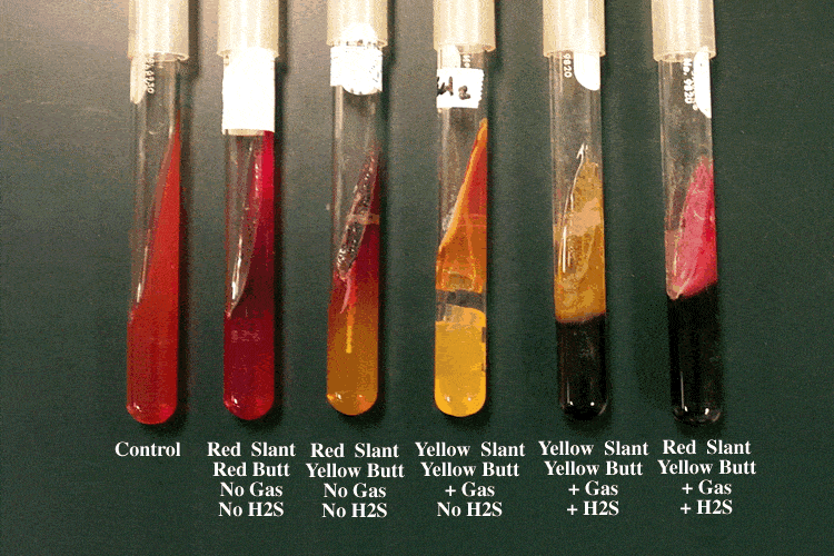
  1. If lactose (or sucrose) is fermented, a large amount of acid is produced, which turns the phenol red indicator yellow both in butt and in the slant. Some organisms generate gases, which produces bubbles/cracks on the medium.
  2. If lactose is not fermented but the small amount of glucose is, the oxygen deficientbutt will be yellow (remember that butt comparatively have more glucose compared to slant i.e. more media more glucose), but on the slant the acid (less acid as media in slant is very less) will be oxidized to carbondioxide and water by the organism and the slant will be red (alkaline or neutral pH).
  3. If neither lactose/sucrose nor glucose is fermented, both the butt and the slant will be red. The slant can become a deeper red-purple (more alkaline) as a result of production of ammonia from the oxidative deamination of amino acids (remember peoptone is a major constitutents of TSI Agar) .
  4. if H2S is produced, the black color of ferrous sulfide is seen.
So the expected results of TSI Agar test are:

TSI Agar Test results
Triple Sugar Iron Agar Test Results
Image source: Clark College

  1. Alkaline slant/no change in butt (K/NC) i.e Red/Red = glucose, lactose and sucrose non-fermenter
  2. Alkaline slant/Alkaline butt (K/K) i.e Red/Red = glucose, lactose and sucrose non-fermenter
  3. Alkaline slant/acidic butt (K/A); Red/Yellow = glucose fermentation only, gas (+ or -), H2s (+ or -)
  4. Acidic slant/acidic butt (A/A); Yellow/Yellow = glucose, lactose and/or sucrose fermenter gas (+ or -), H2s (+ or -).
Some example of Triple Sugar Iron (TSI) Agar Reactions: 

Name of the organismsSlantButtGasH2S
Escherichia, Klebsiella, EnterobacterAcid (A)Acid (A)Pos (+)Neg (-)
Shigella, SerratiaAlkaline (K)Acid (A)Neg (-)Neg (- )
Salmonella, ProteusAlkaline (K)Acid (A)Pos (+)Pos (+)
PseudomonasAlkaline (K)Alkaline (K)Neg (-)Neg (-)

PYR- test Biokimia Bakteri

PYR Test- Principle, Uses, Procedure and Result Interpretation

PYR (Pyrrolidonyl Aminopeptidase) Test is used for the detection of pyrolidonyl arylamidase (also called pyrrolidonyl aminopeptidase) activity in Streptococcus pyogenes (group A strep), Enterococcus spp., some coagulase-negative staphylococci, and some Enterobacteriaceae. It is also known as PYR (L-pyrrolidonyl-β-naphthylamide) which serve as a substrate for the detection of pyrrolidonyl peptidase.
Facklam, Thacker, Fox and Eriquez reported that 98% of group A streptococci and 96% of group D enterococci hydrolyze PYR. Although Aerococcus species are rarely isolated in the clinical laboratory, these organisms are also expected to hydrolyze PYR.
Facklam et al. further reported that 98% of group B streptococci, 100% of non-group A, B and D streptococci, 100% of group D non-enterococci and 82% of viridans streptococci yield negative PYR test results.

Principle of PYR Test

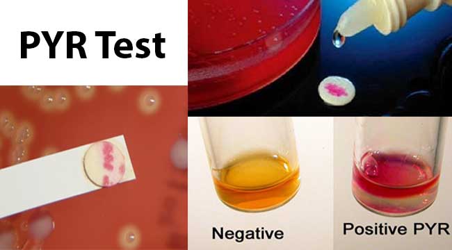
PYR is a rapid method for presumptive identification of bacteria based on the pyrrolidonyl arylamidase enzyme. The enzyme L-pyrrolidonyl arylamidase hydrolyzes the L-pyrrolidonyl- β-naphthylamide substrate to produce a β-naphthylamine. The β-naphthylamine can be detected in the presence of N,N-methylaminocinnamaldehyde reagent by the production of a bright red precipitate.
Following hydrolysis of the substrate by the peptidase, the resulting b-naphthylamide produces a red color upon the addition of 0.01% cinnamaldehyde reagent. When a visible inoculum of microorganism is rubbed onto a small area of a disk impregnated with the substrate, the hydrolysis occurs within 2 min, at which time the cinnamaldehyde reagent is added to detect the reaction by a color change to purple.

Uses of PYR Test

  1. It is used for the presumptive identification of group A streptococci (Streptococcus pyogenes).
  2. It is used for the rapid differentiation of enterococci from group D ß-hemolytic streptococci.
  3. It also differentiate some Staphylococci (positive haemolyticus from negative S. auricularis).
  4. It is used in the identification of E. coli, separating it from other indole positive, lactose positive, gram-negative rods.

Procedure of PYR Test

Broth Method
  1. Inoculate PYR broth with 3-5 colonies from 18-24 hours pure culture.
  2. Incubate the tube aerobically at 35-37°C for 4 hours.
  3. Add 2-3 drop of PYR reagent and observe for color change.
  4. Observe for the red color development within 1-2 minutes.
Disk Method (Rapid)
  1. Wet the PYR test disc on the strip with 10 µl sterile distilled water or deionized water.
Note: Do not flood the disk.
  1. Put 5-10 colonies of the tested strain from 18-24 hours culture on the surface of the disc with a loop and smear them lightly on it.
  2. Incubate the disc for 1-2 minutes at room temperature.
  3. After incubation, add 1 drop of N, N-dimethylaminocinnamaldehyde.
  4. Observe for red color development within 1-2 minutes.

Result Interpretation of PYR Test

Result Interpretation of PYR Test
Positive: Bright pink or cherry-red color within 1-2 minutes.
Examples: Group A Streptococci (Streptococcus pyogenes), Group D Enterococci (Enterococcus faecalis and Enterococcus faecium), Coagulase negative Staphylococcus species such as S. hemolyticus, S. lugdunensis, S. schleiferi., Enterobacter, Citrobacter, Klebsiella, Yersinia and Serratia, Aerococcus, Gamella, Lactococcus, most Corynebacterium (Arcanobacterium) hemolyticum.
Negative: No color change or a blue color due to a positive indole reaction.
Examples: Group B Streptococci (Streptococcus agalactiae), Streptococcus mitis, S. bovis, S. equinus, S. milleri.
Note: A pale pink reaction (weak) is considered negative.

Quality Control for PYR Test

Positive Control: Enterococcus faecalis (ATCC29212), Streptococcus pyogenes(ATCC19615)
Negative Control: Streptococcus agalactiae (ATCC10386)

Limitations of PYR Test

  1. PYR may be used in the presumptive separation of group A streptococci and group D enterococci from other streptococci. Additional testing, using a pure culture, is recommended for complete identification.
  2. A false-negative test can result if the disk or filter paper are too moist.
  3. False-negative tests can result if selective media or tube biochemical agars are used to provide inocula.
  4. Escherichia coli and indole-positive Proteus  obtained from media containing a high tryptophan content may yield a blue-green color development. This is a negative result.
  5. Some less commonly encountered isolates of lactococci and aerococci may be PYRase positive.
  6. Non-specific colour reactions may occur if results are read after 20 seconds.

References

  1. PYR TEST. ITEST PRO. S.R.O. Code: SO 341.
  2. PYR Test Kit by Hardy Diagnostics. Medline Industries, Inc.
  3. PYR Test Kit. Key Diagnostic. Cat. no. HDZ75 / HDZ175
  4. PYR (L-Pyrrolidonyl-b-Naphthylamide) Test. Chapter 3.17.41. Clinical Microbiology Procedures Handbook. Volume 1. Second Edition Update (2007).
  5. L-Pyrrolidonyl Arylamidase (PYR) Test. Procedure 13-36. Bailey & Scott’s Diagnostic Microbiology.
  6. Monica Cheesbrough. District Laboratory Practice in Tropical Countries. Second Edition. Part 2. Chapter 7 Microbiological Test. 7.18.2 Streptococcus pyogenes. pp- 160.
  7. Patrick R Murray. Manual of Clinical Microbiology. 8th PRY Test. pp- 409-410.
  8. PYR Disk. Remel.
  9. PYR Test Kit and PYR Reagent. Hardy Diagnostics.
  10. PYR Test. Procedure Manual Toronto Medical Laboratories / Mount Sinai Hospital Microbiology Department.
  11. PYR Test Kit and PYR Reagent. CONDA Lab.
  12. Prospot™ PYR Test Kit. Pro-Lab Diagnostics.
  13. Mehdi Aslani and Reza Gharagozloo. 1995. Rapid Identification of Group A Streptococci by Pyrrolidonyl-B-Naphthylamide Hydrolysis. Medical Journal of the Islamic Republic of Iran. 9(3):243-245.
  14. Facklam RR, Thacker LG, Fox B and Eriquez L. 1982. Presumptive identification of streptococci with a new test system. J. Clin. Microbiol. 15: 987-990.
  15. Oberhofer TR. 1986. Value of the L-pyrrolidonyl-b-napthylamide hydrolysis test for identification of select gram-positive cocci. Diagn. Microbiol. Infect. Dis. 4: 43-47.
  16. Ruoff KL. 2003. Aerococcus, Abiotrophia and other infrequently isolated aerobic catalase-negative, gram-positive cocci. In Murray, P.R., E.J. Baron, J.H. Jorgensen, M.A. Pfaller and R.H. Yolken (ed.), Manual of clinical microbiology, 8th ed., Vol. 1. American Society for Microbiology, Washington, D.C.
  17. http://microbeonline.com/pyrrolidonyl-arylamidase-pyr-test-principle-procedure-results/
  18. http://www.microbiologynotes.com/pyr-l-pyrrolidonyl-β-naphthylamide-test-principle-uses-procedure-result-interpretaion-examples-and-limitation/
  19. http://www.microbiologyinpictures.com/bacteria-photos/enterococcus-faecalis-images/enterococcus-faecalis-pyr-test.html
  20. http://www.bacteriainphotos.com/pyr_test.html

media nutrien agar

Nutrient Agar: Composition, Preparation and Uses

Nutrient Agar is a general purpose, nutrient medium used for the cultivation of microbes supporting growth of a wide range of non-fastidious organisms. Nutrient agar is popular because it can grow a variety of types of bacteria and fungi, and contains many nutrients needed for the bacterial growth.
Composition of Nutrient Agar
  • 0.5% Peptone

It is an enzymatic digest of animal protein. Peptone is the principal source of organic nitrogen for the growing bacteria.
  • 0.3% beef extract/yeast extract

It is the water-soluble substances which aid in bacterial growth, such as vitamins, carbohydrates, organic nitrogen compounds and salts.
  • 1.5% agar

It is the solidifying agent.
  • 0.5% NaCl

The presence of sodium chloride in nutrient agar maintains a salt concentration in the medium that is similar to the cytoplasm of the microorganisms.
  • Distilled water

Water is essential for the growth of and reproduction of micro-organisms and also provides the medium through which various nutrients can be transported.
  • pH is adjusted to neutral (7.4) at 25 °C.

Preparation of Nutrient Agar
1. Suspend 28 g of nutrient agar powder in 1 litre of distilled water.
2. Heat this mixture while stirring to fully dissolve all components.
3. Autoclave the dissolved mixture at 121 degrees Celsius for 15 minutes.
4. Once the nutrient agar has been autoclaved, allow it to cool but not solidify.
5. Pour nutrient agar into each plate and leave plates on the sterile surface until the agar has solidified.
6. Replace the lid of each Petri dish and store the plates in a refrigerator.
Uses of Nutrients Agar
1. It is frequently used for isolation and purification of cultures.
2. It can also be used as a means for producing the bacterial lawns needed for antibiotic sensitivity tests.  In actuality, antibiotic sensitivity testing is typically performed on media specially formulated for that purpose.
Pictures
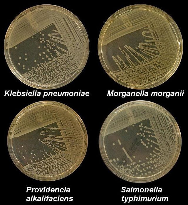
Four nutrient agar plates growing colonies of common Gram negative bacteria.
Source: Wikipedia
Staphylococcus aureus in nutrient agar
Staphylococcus aureus in nutrient agar.
Source: CHARLES UNIVERSITY
Staphylococcus epidermidis in nutrient agar.
Staphylococcus epidermidis in nutrient agar.                Source: CHARLES UNIVERSITY

MEDIA UJI BIOKIMIA BAKTERI

Simmons Citrate Agar- Composition, Principle, Uses, Preparation and Result Interpretation

Simmons Citrate Agar is an agar medium used for the differentiation of Enterobacteriaceae based on the utilization of citrate as the sole source of carbon. 
In the early 1920s, Koser developed a liquid medium formulation for the differentiation of fecal coliforms from the coliform group. Simmons later modified this formulation to produce a solid medium that eliminated potential errors when interpreting growth.

Composition of Simmons Citrate Agar

Ingredients per liter of deionized water:*
Sodium Chloride (NaCl)5.0 gm
Sodium Citrate (dehydrate)2.0 gm
Ammonium Dihydrogen Phosphate1.0 gm
Dipotassium Phosphate1.0 gm
Magnesium Sulfate (heptahydrate)0.2 gm
Bromothymol Blue0.08 gm
Agar15.0 gm
Deionized water = 1,000 ml
Final pH 6.9 +/- 0.2 at 25 degrees C.

Principle of Simmons Citrate Agar

Simmons Citrate agar is used to test an organism’s ability to utilize citrate as a source of energy. Ammonium Dihydrogen Phosphate is the sole source of nitrogenDipotassium Phosphate acts as a bufferSodium Chloride maintains the osmotic balance of the medium. Sodium Citrate is the sole source of carbon in this medium. Magnesium Sulfate is a cofactor for a variety of metabolic reactions. Bacteriological agar is the solidifying agent. Organisms capable of utilizing ammonium dihydrogen phosphate and citrate will grow unrestricted on this medium. If citrate can be used, the microbe will accumulate alkaline/basic byproducts.
Bacteria that can grow on this medium produce an enzyme, citrate-permease, capable of converting citrate to pyruvatePyruvate can then enter the organism’s metabolic cycle for the production of energy. Growth is indicative of utilization of citrate, an intermediate metabolite in the Krebs cycle.
When the bacteria metabolize citrate, the ammonium salts are broken down to ammonia, which increases alkalinity. The shift in pH turns the bromthymol blueindicator in the medium from green to blue above pH 7.6.

Uses of Simmons Citrate Agar

  1. It is used for the differentiation of Gram-negative bacteria on the basis of citrate utilization.
  2. Simmons Citrate Agar may be used to differentiate citrate-positive Salmonella enteritidis and members of Salmonella subgenus II, III and IV from the citrate-negative Salmonella typhi, Salmonella paratyphi A, Salmonella pullorum and Salmonella gallinarum.
  3. Simmons Citrate Agar is primarily used to aid in the identification of Enterobacteriaceae. Uses
    include:
  • Escherichia coli (usually -) and Shigella spp. (-) from other commonly encountered
    Enterobacteriaceae (variable +).
  • Edwardsiella spp. (-) from Salmonella spp. (usually +)
  • Serratia proteamaculans (+) from Yersinia pseudotuberculosis (-) and Yersinia enterocolitica
    (usually -)
  • Klebsiella-Enterobacter groups (usually +) from E. coli (usually -)
  • Proteus rettgeri (+) from Morganella morganii biogroups 1 and 2 (-)
  • Yokenella regensburgei (+) from Hafnia alvei (-)
  • Leminorella grimontii (+) from L. richardii (-)
  • Acidovorax delafieldii (+) from A. facilis (-) and  A. temperans (-)

Preparation of Simmons Citrate Agar

  1. Dissolve above salts in deionized water.
  2. Adjust pH to 6.9.
  3. Add agar and Bromothymol blue.
  4. Gently heat, with mixing, to boiling until agar is dissolved.
  5. The medium may be used either as slopes in test tubes or as a plate medium in petri dishes. In both cases the surface of the medium is lightly inoculated by streaking and, where slopes are used, the butt of medium is inoculated by stabbing.
  6. For tubes, dispense 4.0 to 5.0 ml into 16-mm tubes.
  7. Autoclave at 121 degree C under 15 psi pressure for 15 minutes.
  8. Cool in slanted position (long slant, shallow butt).
  9. Tubes should be stored in a refrigerator to ensure a shelf life of 6 to 8 weeks.
  10. The uninoculated medium will be a deep forest green due to the pH of the sample and the bromothymol blue.

Result Interpretation on Simmons Citrate Agar

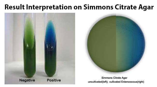
Positive growth (i.e. citrate utilisation) produces an alkaline reaction and changes the colour of the medium from green to bright blue.
Examples: Serratia and the majority of the Enterobacter, Citrobacter, Klebsiella, Proteus and Providencia species, except Morganella morganii and Klebsiella rhinoscleromatis.
Negative test (i.e. no citrate utilisation) the colour of the medium remains unchanged.
Examples: Escherichia coli, Shigella, Yersinia, Edwardsiella species, etc.

Result Interpretation on Simmons Citrate Agar

Limitations of Simmons Citrate Agar

  1. It is important not to carry over any nutrients into the citrate medium because this will result in false positive tests.
  2. Some organisms are capable of growth on citrate and do not produce a color change. Growth is considered a positive citrate utilization test, even in the absence of a color change.
  3. Tests with equivocal results should be repeated.
  4. The reactions of this medium alone are not sufficient for identification to the species level.
  5. The inoculum should be from a pure, overnight culture grown on a solid medium and not from a broth suspension.

Quality Control on Simmons Citrate Agar

Positive control: Klebsiella pneumoniae ATCC® 13883. Growth; colour change of medium to blue 
Negative control: Escherichia coli ATCC® 25922. Inhibited or no growth; no colour change of medium

References

  1. Thermo Fisher Scientific Inc., Dehydrated Culture Media- SIMMONS CITRATE AGAR.
  2. Sisco Research Laboratories Pvt. Ltd.- Simmon’s Citrate Agar (59437).
  3. Austin Community College.
  4. Hardy Diagnostics- SIMMONS CITRATE AGAR (L80).
  5. Intuitive Systems, Inc.- Simmons’ Citrate Agar Slant.
  6. Scharlab- Simmons Citrate Agar (01-177).
  7. Merck Microbiology Manual 12th Edition- SIMMONS Citrate Agar.
  8. Dalynn Biologicals- SIMMONS CITRATE AGAR (TS62).
  9. Remel- Citrate Agar (Simmons).
  10. Conda Lab- SIMMONS CITRATE AGAR ISO 10273 (1014).
  11. Sigma-Aldrich, Inc.- SIMMONS CITRATE AGAR (S3681).
  12. HiMedia Laboratories Pvt. Ltd.- Simmons Citrate Agar (M099).
  13. Acumedia Manufacturers, Inc.- SIMMONS CITRATE AGAR (7156).

germinasi Candida albicans

tugas pendahuluan mikologi d4 1. jelaskan dan gambarkan perbedaan ciri-ciri species Tricophyton sp, epidermophyton sp , mikrospor...